

Plastination Center
What is Plastination?
Plastination is a chemical process developed to preserve various types of human and animal tissue. "The plastination process works by removing the water from cells and replacing it with acetone. After that, the organ is placed in pure liquid silicone under a vacuum, which causes the acetone to bubble out of the cells and allows the silicone to be sucked into them. The process, which takes about six months from start to finish, results in organs that are not plastic-coated, but are 100 percent plastic throughout." This yields a specimen that is durable, odor free, and anatomically intact.
Plastination is a technique by which specimens are preserved in plastic, without destroying the composition and structure of the tissue. Plastinated specimens can be used as models and teaching tools for students in any field that requires gross anatomical studies. They can also be used for comparing anatomy to imaging such as computed tomography (CT) or magnetic resonance imaging (MRI). Plastination permits the preservation of organs or specimens with normal pathology as well as those with unusual morphology or exhibiting a rare disease. Plastination allows capturing and preserving the disease process and therefore enhances the learning experience.
Why Plastination?
Plastinated specimens offer several advantages over other methods of preservation:
- Enhances the learning experience.
- Specimens are anatomically true, cleaner, drier, and easier to handle.
- Can be used to compare anatomy to MRI and CT analysis.
- Specimens are odorless, more durable, and free from encasing material.
- Minimizes handling of embalmed biological specimens which contain hazardous preservatives.
- Typically retain the natural contours of the specimen.
- Specimens can be used repeatedly — course after course.
Unionite Article
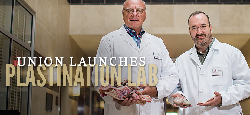
June 2017 - For more than a decade, Union science and nursing students have benefited from their study of human anatomy thanks to a cadaver lab and the generosity of donors who have bequeathed their bodies to Union for scientific study.
Now, Union students can take advantage of a new development — a plastination lab.
Why is the Plastination Lab beneficial to Union?
The plastination laboratory sets Union apart from other universities teaching gross anatomy. Before Union acquired the plastination laboratory, only major medical teaching institutions had this capability. It is estimated that only 400 plastination facilities exist worldwide. The University of Tennessee of Knoxville is the only recognized laboratory in the southeast.
The process was developed in Germany in 1978 and is more prevalent worldwide than it is in the United States. Because many other countries don't have the legal restrictions that exist in the United States, it's easier for the process to be commercialized and profitable.

Union University's plastination lab, which was opened January of 2017, was the eighth plastination lab in the United States. However, due to so few people being trained to work in these labs, some of those eight have become inactive.
"Students in Union's Certified Registered Nurse Anesthetist and College of Pharmacy programs do full cadaver dissections each year, a process that takes about 60 hours. As they dissect the cadaver, students discuss pathology and discover diseased organs. Those organs are then harvested (with permission) and preserved in formaldehyde for future teaching purposes. Even in formaldehyde, however, the organs deteriorate over time. Also, so students are not directly exposed to formaldehyde as they handle the organs, the specimens must go through a time-consuming water bath process prior to the class."
"The university's current donor list of anatomical bequests is about 100 people. Union takes those donations seriously, because the human body is God's creation. The development of the plastination lab will not change how the university handles those donations. It's a very special gift that they give," he says. "We make sure that we're handling them with dignity and respect."
Lab Origins and Ways You Can Get Involved
Dr. Bob Henry, the most experienced plastinator in the US, visited our campus and helped set up our facility. He set up the University of Michigan, University of Toledo Medical Center, and the University of Tennessee labs. His Christian faith was the motivating force in helping Union make this become an successful endeavor.
Now we are attempting to raise enough money to open a museum that would present and display some of the plastinated specimens, so area grade school and high school students could learn from Union's efforts as well. If you feel led to help us with this endeavor, all donations are appreciated.
Donate Now Anatomical Bequests
Recent Plastination Scholarship from Union
- "Application of Pigments to Squirrel Legs at Various Stages of the Plastination Process,", R. Lewis, J.A. Huggins, R.A. Wamble, M.G. Bolyard. Journal of Plastination. December 2020
- "Analysis of Radio Frequency Identification Tagging of Biological Specimens Prior to Plastination" G. R. Vandezande, R.A. Wamble, J.A. Huggins, J.R. Kerfoot, and M. G. Bolyard. Journal of Plastination, 31(2): 6-14, 2019.